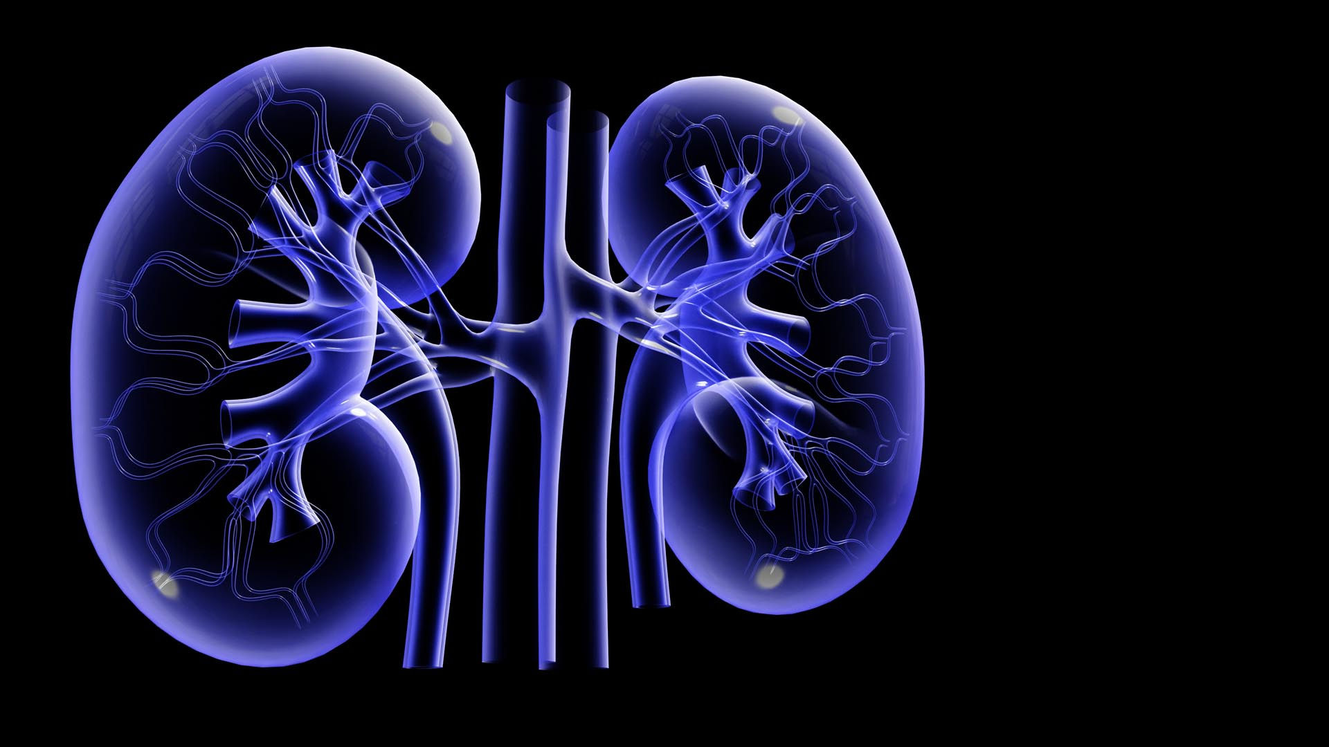
Position
The kidneys are a pair of reddish organs shaped like kidney beans. They lie on either side of the vertebral column between the peritoneum and the back wall of the abdominal cavity at the level of the 12th thoracic and first three lumbar vertebrae. The 11th and 12th pairs of ribs provide some protection for the superior parts of the kidneys. The right kidney is slightly lower than the left because the liver occupies a large area above the kidney on the right side.
External Anatomy
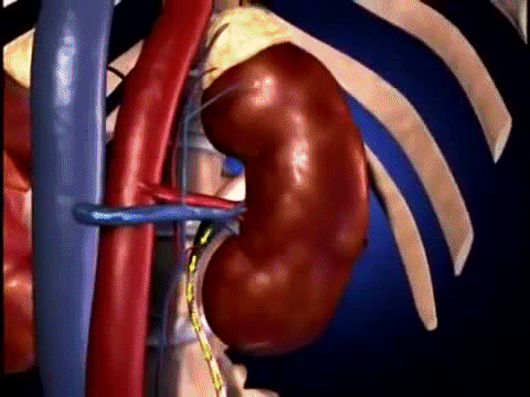
Near the center of the medial border is an indentation called the
renal hilum, through which the ureter leaves the kidney, and blood
vessels, lymphatic vessels, and nerves enter and exit.
Surrounding each kidney is the smooth, transparent renal capsule,
a connective tissue sheath that helps maintain the shape of the
kidney and serves as a barrier against trauma .
Adipose (fatty) tissue surrounds the renal capsule and cushions
the kidney. Along with a thin layer of dense irregular connective
tissue, the adipose tissue anchors the kidney to
the posterior abdominal wall.
Internal Anatomy
Internally, the kidneys have two main regions: an outer lightred
region called the renal cortex (cortex rind or back) and
an inner, darker red-brown region called the renal medulla
(medulla inner portion). Within the renal medulla are several
cone-shaped renal pyramids. Extensions of the renal cortex, called
renal columns, fill the spaces between renal pyramids.
Urine formed in the kidney drains into a large, funnelshaped
cavity called the renal pelvis. The rim of the renal pelvis contains
cuplike structures called major and minor calyces.
Urine flows from several ducts within the kidney into a minor
calyx and from there through a major calyx into the renal
pelvis, which connects to a ureter. Water and solutes in the
fluid that drains into the renal pelvis remain in the urine and
are excreted.

The Kidneys
The Urinary System
The urinary system consists of two kidneys, two ureters, a urinary bladder and a urethra. Nephrology is the study of the anatomy, physiology and disorders of kidney.


About 20–25 percent of the resting cardiac output—1200 milliliters of blood per minute—flows into the kidneys through the right and left renal arteries. Within each kidney, the renal artery divides into smaller and smaller vessels (segmental, interlobar, arcuate, cortical radiate arteries) that eventually deliver blood to the afferent arterioles (af- toward; -ferre to carry). Each afferent arteriole divides into a tangled capillary network called a glomerulus. The capillaries of the glomerulus reunite to form an
efferent arteriole. Upon leaving the glomerulus, each efferent arteriole divides to form a network of capillaries around the kidney tubules (described shortly). These peritubular capillaries eventually reunite to form peritubular veins, which merge into cortical radiate, arcuate, and interlobar veins. Ultimately, all of these smaller veins drain into the renal vein.

The Nephron
The functional units of the kidney are called the nephrons, numbering about a million in each kidney. A nephron consists of two parts: a renal corpuscle, where blood plasma is filtered, and a renal tubule into which the filtered fluid, called glomerular filtrate, passes. Closely associated with a nephron is its blood supply. As the fluid moves through the renal tubules, wastes and excess substances are added, and useful materials are returned to the blood in the peritubular capillaries.
The two parts that make up a renal corpuscle are the
glomerulus and the glomerular (Bowman’s) capsule, a
double-walled cup of epithelial cells that surrounds the glomerular capillaries. Glomerular filtrate first enters the glomerular capsule and then passes into the renal tubule. In the order that fluid passes through them, the three main sections of the renal tubule are the proximal convoluted tubule, the nephron loop, and the distal convoluted tubule. Proximal denotes
the part of the tubule attached to the glomerular capsule, and distal denotes the part that is farther away. The renal corpuscle and both convoluted tubules lie within the renal cortex; the nephron loop extends into the renal medulla. The first part
of the nephron loop begins at the point where the proximal convoluted tubule takes its final turn downward. It begins in the renal cortex and extends downward into the renal medulla and
is called the descending limb of the nephron loop.
It then makes a hairpin turn and returns to the renal cortex where it terminates at the distal convoluted tubule and is known as the ascending limb of the nephron loop. The distal convoluted tubules of several nephrons empty into a common collecting duct.


The Ureters, Bladder and Urethra
The Ureter
Each of the two ureters transports urine from the renal pelvis of one of the kidneys to the urinary bladder. The ureters pass under the urinary bladder for several centimeters, causing the bladder to compress the ureters and thus prevent backflow of urine when pressure builds up in the bladder during urination. The wall of the ureter consists of three layers. The inner layer is the mucosa, containing transitional epithelium with an underlying layer of areolar connective tissue.
Transitional epithelium is able to stretch—a marked advantage for any organ that must accommodate a variable volume of fluid. Mucus secreted by the goblet cells of the mucosa prevents the cells from coming in contact with urine, the solute concentration and pH of which may differ drastically from the cytosol of cells that form the wall of the ureters. The middle layer consists of smooth muscle. Urine is transported from the renal pelvis to the urinary bladder primarily by peristaltic contractions of this smooth muscle, but the fluid pressure of the urine and gravity may also contribute. The outer layer consists of areolar connective tissue containing blood vessels, lymphatic vessels, and nerves.
The Urinary Bladder
The urinary bladder is a hollow muscular organ situated in the pelvic cavity behind the pubic symphysis. Folds of the peritoneum hold the urinary bladder in position. The shape of the urinary bladder depends on how much urine it contains. When empty, it looks like a deflated balloon. It becomes spherical when slightly stretched and, as urine volume increases, becomes pear-shaped and rises into the abdominal cavity. Urinary bladder capacity averages 700–800mL. It is smaller in females because the uterus occupies the space just superior to the urinary bladder. Toward the base of the urinary bladder, the ureters drain into the urinary bladder via the ureteral openings. Like the ureters, the mucosa of the urinary bladder contains transitional epithelium, which permits stretching. The mucosa also contains folds called rugae, which also permit expansion of the urinary bladder. The muscular layer of the urinary bladder wall consists of three layers of smooth muscle called the detrusor muscle. The peritoneum, which covers the superior surface of the urinary bladder, forms a serous outer coat; the rest of the urinary bladder has a fibrous outer covering.
The Urethra
The urethra, the terminal portion of the urinary system, is a small tube leading from the floor of the urinary bladder to the exterior of the body. In females, it lies directly behind the pubic symphysis and is embedded in the front wall of the vagina. The opening of the urethra to the exterior, the external urethral orifice, lies between the clitoris and vaginal opening. In males, the urethra passes vertically through the prostate, the deep perineal muscles, and finally the penis. Around the opening to the urethra is an internal urethral sphincter composed of smooth muscle. The opening and closing of the internal urethral sphincter is involuntary. Below the internal sphincter is the external urethral sphincter, which is composed of skeletal muscle and is under voluntary control.
